What Does Breast Cancer Look Like On Imaging / Ai Tool Improves Breast Cancer Detection On Mammography Imaging Technology News / Any area that does not look like normal tissue is a possible cause for concern.. A diagnostic mammogram is used to check for breast cancer when there is a sign or symptom of disease, or to evaluate an abnormality seen on a screening mammogram. A breast mri usually is performed after you have a. The limitations of mammography in the detection and evaluation of invasive lobular carcinoma (ilc) have long been recognized, presenting real clinical challenges in treatment planning for these tumors. A picture is worth a thousand words. To help determine the extent of breast cancer:
Other ultrasound findings that suggest breast cancer include: But not every woman who has been diagnosed with breast cancer needs a breast mri. A breast mri captures multiple images of your breast. It also can be used to look at a suspicious area that was seen on a mammogram. Lobular breast cancer can be more difficult to see on imaging and scans.
Follow Up Of Patients With Breast Cancer Imaging Of Local Recurrence And Distant Metastases Springerlink from media.springernature.com There are different kinds of asymmetries, from difference in size to tissue density. What does breast cancer look like? This type of cancer precedes the development of a distinct mass, lump or invasive cancer. The dye collection in the breast can also look clumpy or appear in a section of the breast, depending on the involvement of dcis. The breast tissue kind of looks like waves on the ocean. Imaging and lobular breast cancer. Scintimammography can detect breast cancer even when dense breast tissue or breast implants are present. Both are features we look at on your breast imaging study.
Screening mammograms have been performed since the 1970s.
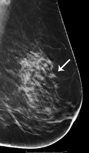
Breast imaging specialists like dr. What does breast cancer look like? A lump or tumor will show up as a focused white area on a mammogram. Baker determine if calcifications are worrisome by looking at the size, shape, and distribution of the flecks, and at any associated mass that might appear in the breast tissue at the same time. A 3d mammogram is used as a breast cancer screening test to look for breast cancer in people with no signs or symptoms of the disease. These images are called mammograms. The person operating the ultrasound will sweep, or fan, the probe back and forth to look at different areas in 90 degree angle images. The limitations of mammography in the detection and evaluation of invasive lobular carcinoma (ilc) have long been recognized, presenting real clinical challenges in treatment planning for these tumors. As with all abnormalities seen on breast imaging, the diagnosis of dcis requires a sample of tissue or biopsy. Any area that does not look like normal tissue is a possible cause for concern. Lobular breast cancer can be more difficult to see on imaging and scans. Scintimammography can detect breast cancer even when dense breast tissue or breast implants are present. Before the test, you may need to have a contrast solution (dye) injected into your arm through an intravenous line.
As with all abnormalities seen on breast imaging, the diagnosis of dcis requires a sample of tissue or biopsy. Scintimammography can detect breast cancer even when dense breast tissue or breast implants are present. This is considered an abnormal mammogram, but not necessarily one that's indicative of cancer. It also can be used to look at a suspicious area that was seen on a mammogram. Before the test, you may need to have a contrast solution (dye) injected into your arm through an intravenous line.
Breast Cancer Screening With 3d Mammography Or Tomosynthesis Radiology Imaging Ma Ct from www.rad-imaging.com A breast mri (magnetic resonance imaging) is a test that is sometimes performed along with a screening mammogram in women with at least a 20% lifetime risk of developing breast cancer. Learn about getting a mammogram. Before the test, you may need to have a contrast solution (dye) injected into your arm through an intravenous line. But radiologists can still see signs of cancer. But not every woman who has been diagnosed with breast cancer needs a breast mri. This is considered an abnormal mammogram, but not necessarily one that's indicative of cancer. Ultrasound is useful for looking at some breast changes, such as lumps (especially those that can be felt but not seen on a mammogram) or changes in women with dense breast tissue. Calcifications are tiny flecks of calcium — like grains of salt — in the soft tissue of the breast that can sometimes indicate the presence of an early breast cancer.
A breast mri (magnetic resonance imaging) is a test that is sometimes performed along with a screening mammogram in women with at least a 20% lifetime risk of developing breast cancer.
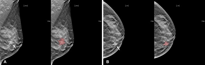
Any area that does not look like normal tissue is a possible cause for concern. Dense breast tissue appears solid. It can also be used to investigate breast problems, such as a suspicious lump or thickening. Magnetic resonance imaging (mri) of the breast — or breast mri — is a test used to detect breast cancer and other abnormalities in the breast. Imaging and lobular breast cancer. It also can be used to look at a suspicious area that was seen on a mammogram. A 3d mammogram is used as a breast cancer screening test to look for breast cancer in people with no signs or symptoms of the disease. However, advances in mammography, ultrasound, and magnetic resonance imaging present opportunities to improve the diagnosis and preoperative assessment of ilc. Lobular breast cancer can be more difficult to see on imaging and scans. This is acceptable for diagnostic exams. A lump or tumor will show up as a focused white area on a mammogram. In this mammogram image, the breast calcifications are in ductal patterns. Breast imaging specialists like dr.
While they're looking for possible cancer, your doctors may also come across masses or structures in the breast that deserve further investigation, including: Both are features we look at on your breast imaging study. The breast tissue kind of looks like waves on the ocean. Baker determine if calcifications are worrisome by looking at the size, shape, and distribution of the flecks, and at any associated mass that might appear in the breast tissue at the same time. Finding breast lumps and seeing change in the size and shape.
Prediction Of Breast Cancer Molecular Subtypes Using Radiomics Signatures Of Synthetic Mammography From Digital Breast Tomosynthesis Scientific Reports from media.springernature.com Both are features we look at on your breast imaging study. However, in rare cases, breast cancer can be the cause of gynecomastia so, a full mammographic. In this mammogram image, the breast calcifications are in ductal patterns. On a mammogram, an asymmetry typically means there's more tissue, or white stuff on the mammogram, in one area than on the opposite side. What does a cancerous breast lump look like on ultrasound? This is considered an abnormal mammogram, but not necessarily one that's indicative of cancer. A diagnostic mammogram is used to check for breast cancer when there is a sign or symptom of disease, or to evaluate an abnormality seen on a screening mammogram. On ultrasound, a breast cancer tumor is often seen as hypoechoic, has irregular borders, and may appear spiculated.
The limitations of mammography in the detection and evaluation of invasive lobular carcinoma (ilc) have long been recognized, presenting real clinical challenges in treatment planning for these tumors.
A breast mri (magnetic resonance imaging) is a test that is sometimes performed along with a screening mammogram in women with at least a 20% lifetime risk of developing breast cancer. Finding breast lumps and seeing change in the size and shape. There are different kinds of asymmetries, from difference in size to tissue density. Ductal carcinoma in situ is usually seen as linear microcalcifications (see arrows), which demonstrate linear orientation, as seen in this patient. A breast mri usually is performed after you have a. This type of cancer precedes the development of a distinct mass, lump or invasive cancer. A screening mammogram is performed at regular intervals to check for breast cancer in women who have no signs or symptoms of the disease. The person operating the ultrasound will sweep, or fan, the probe back and forth to look at different areas in 90 degree angle images. These images are called mammograms. On a mammogram, an asymmetry typically means there's more tissue, or white stuff on the mammogram, in one area than on the opposite side. Imaging and lobular breast cancer. Breast mri is sometimes used in women who already have been diagnosed with breast cancer, to help measure the size of the cancer, look for other tumors in the breast, and to check for tumors in the opposite breast. Breast imaging specialists like dr.